Return to QEQM main Display
Display Contents
QEQM Hospital Margate
Link - The skeletons
The Beaker burial
Condition
Sex
Age
The skull
The spine
The pelvis
The arm bones
The leg bones
The secondary burial
Condition
Sex
Age
Stature
The skull
The pelvis
The arm bones
The leg bones
Link - The skeletons
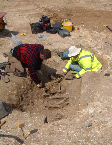
Photo by TTA
Photo by Susan Deacon (TTA)
Small scale in centimetre divisions
Large scale in 10 centimetre divisions
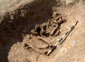
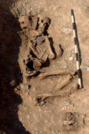
The QEQM Beaker burial
Photo by Susan Deacon (TTA)
Scale in centimetre divisions
Sarah Tatham reported that
the burial showed extensive
surface erosion and
fragmentation, though was remarkably complete considering the length of
inhumation. All areas were well represented with the exception of hands
and feet and some crushed parts of the skull.
Stature could not be reliably determined however.
Stature could not be reliably determined however.
Photo by Susan Deacon (TTA)
Small scale in centimetre divisions
Large scale in 10 centimetre divisions
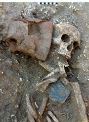
Chew Beaker
Photo by TTA
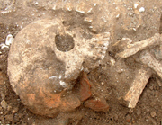
Tooth wear was commensurate
with an individual of 25-35 years of age
(Brothwell 1981), but this is an unreliable age marker and more likely
indicated that his diet did not require the excessive grinding of his
teeth. The upper maxilla had complete dentition (including wisdom
teeth) and no signs of disease.
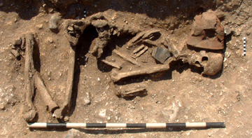
The QEQM Beaker burial
Photo by Susan Deacon (TTA)
Small scale in centimetre divisions
Large scale in 10 centimetre divisions
The vertebrae showed much
pitting and age-related changes, with
extensive osteophytes (and notably the beginnings of kyphosis) present
in the last two lumbar and first sacral vertebrae).
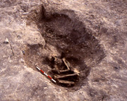
Photo by TTA
Scale in 10 centimetre divisions
at QEQM
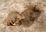
The rectangular shape of the Beaker grave is actually the part-excavated fill of the proposed coffin-structure
This is surrounded by a chalk backfill which blends into the natural chalk beyond, making the large oval shape of the grave cut hard to see.
Photo by TTA
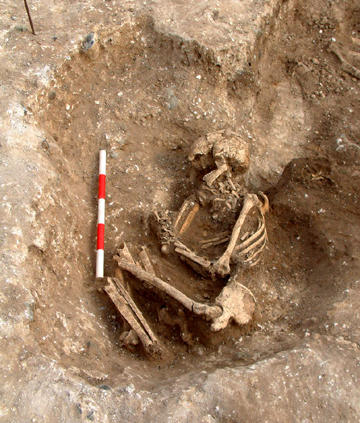
Condition
As with the Beaker burial, this skeleton also showed extensive surface erosion and fragmentation, though was remarkably complete considering the length of inhumation. All areas were well represented with the exception of hands and feet and some crushed parts of the skull.
No evidence of disease or injury had survived post-mortem bone erosion.
Sex
The bones were those of an adult female.
The left humerus (arm bone)
was the only complete bone of the
skeleton and permitted an estimate of the maximum stature of the
individual as 1.59m.
Photo by TTA
Scale in 10 centimetre divisions

The mandible (lower jaw)
was almost complete and the teeth
free of disease, though the presence of some calculus (plaque) may have
caused gum disease. Dental attrition suggested an age of 17-25 years
(Brothwell 1981), though this wear is also related to diet.
Evidence from the pelvis
suggested that this bone was free of injury and disease.
The secondary burial at QEQM
Photo by TTA
The left humerus (upper arm
bone) had well defined muscle markers and gave
evidence of an active life. It was the only complete bone of the
skeleton.
The right clavicle (shoulder) also showed evidence of strong muscle attachments.
The right clavicle (shoulder) also showed evidence of strong muscle attachments.
The diaphysis of the left
tibia and
fibula (lower leg bones) showed high levels of muscle use.
TTA - Trust for Thanet Archaeology.
Brothwell D. 1981. (Precise details unknown).
Tatham S. 2006. The Human Bone in Gardner O.W. and Moody G.A. Queen Elizabeth the Queen Mother Hospital, St. Peter’s Road, Margate, Kent. Trust for Thanet Archaeology report, Part 4.
Much thanks goes to Dr. Sarah Tatham for her analysis of the skeletons.
Thanks also to John Villette for the use of the 1 metre black and white photographic scale.
Version 1 - Posted 16.12.06
All
content © Trust for Thanet Archaeology